
Sex-Specific Risk Factors of Pancreatic Cancer
Petra Samardzija, BMSc
Contact: petrasam3@gmail.com
November 14, 2020
In order to develop more targeted diagnostic and treatment approaches for cancer, it is important to understand the sex-specific variations that exist in cancer development. Specifically, these differences must be considered in one of the most prevalent forms of pancreatic cancer, called pancreatic ductal adenocarcinoma (PDAC). PDAC is the fourth most common cause of cancer-related deaths worldwide and has a 5-year survival rate of only 9%. Difficulty diagnosing the cancer in its early stage and poor response to conventional chemotherapeutics contribute to the lethality of PDAC. A research team from the Lawson Health Research Institute and Western University’s Schulich School of Medicine & Dentistry performed a study that was the first to identify a sex-specific genetic risk factor for PDAC. Their article aims to discover a new potential therapeutic target for pancreatic cancer and focuses on the role of two genes, Kras and Atrx, in the development of PDAC.
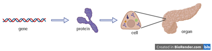
Genes are made up of a molecular structure called DNA and are passed down from generation to generation. They carry instructions to make proteins, which do most of the work in our cells. The type and function of a cell are in part determined by the different proteins that it contains. Millions of cells collectively make up an organ (Figure 1). In cancer, certain gene changes (called mutations) can result in an increased activity of the protein, a loss of protein function, or a protein with an altered function. These changes cause cells to act abnormally and can ultimately disrupt the function of the organ.
The pancreas is a long, flat gland located in the upper abdomen, behind the stomach. It is divided into two major functions: exocrine and endocrine. The endocrine portion of the pancreas is important for controlling blood sugar levels. About 95% of the pancreas is exocrine, which is composed mainly of acinar cells. Pancreatic acinar cells produce enzymes that are released in a tube-like structure called a duct, which connects to the small intestine. Once the enzymes reach the intestine, they help further break down food into smaller molecules, making it easier for the body to absorb the nutrients.
Pancreatitis is a disease in which the pancreas becomes inflamed. Inflammation occurs typically in response to a foreign body (e.g., bacteria, virus, irritant) and acts to remove or “destroy” the “intruder”. However, excessive inflammation can start to cause damage to healthy organs. Chronic pancreatitis (inflammation of the pancreas persisting for a long time) has been shown to increase the risk of pancreatic cancer.
Changes in a gene called Kras that result in the constant activity of the KRAS protein also increase an individual’s susceptibility to pancreatic cancer and is present in 97% of PDAC cases. However, this alteration alone is not enough to cause PDAC, and additional changes in other genes are required.
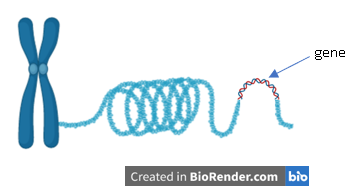
Genes are organized in thread-like strands called chromosomes (Figure 2). Chromosomes can be coiled or uncoiled (“remodelled”) in various ways by chromatin remodeling proteins. The coiling of chromosomes at certain locations influences whether a gene is “expressed” (able to make protein). Previous studies have found that mutations in chromatin remodeling proteins are commonly found in PDAC tumours.
This article was particularly interested in the Atrx gene, which produces the chromatin remodeling protein, ATRX. In a mouse model containing a pancreas lacking the ATRX protein, the ability to repair DNA damage was reduced compared to mice with ATRX. This led researchers to examine the effect of the loss of ATRX in pancreatic injury (such as pancreatitis), where intact DNA repair mechanisms are required for the pancreas to heal. It was hypothesized that a loss in ATRX would enhance the damaging effects of pancreatitis.
To study this, scientists induced pancreatitis using the drug cerulein in male and female mice that either had the ATRX protein or did not have ATRX. Cerulein stimulates the secretion and early activation of pancreatic enzymes, causing damage to the pancreas and eventually pancreatitis. Increased pancreatic damage was seen in the mice lacking ATRX. What caught scientists by surprise was that pancreatic damage was more pronounced in female mice lacking the ATRX protein than in male mice lacking ATRX.
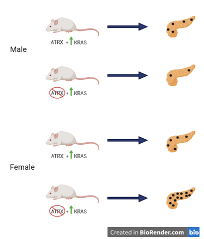
Scientists then proceeded to make another mouse model containing an increased activity of KRAS and no ATRX protein. It was predicted that since the lack of ATRX has been shown to increase the extent of pancreatic injury and increased activity of KRAS is seen in most PDAC cases, the two alterations would result in a damaged pancreas that is seen in cancer. Surprisingly, in male mice, a decrease in damage was seen in the absence of ATRX and the increased activity of KRAS compared to male mice with ATRX present (Figure 3). The greatest amount of damage was observed in female mice lacking ATRX that had increased activity of KRAS.
To translate these findings to humans, the team used the International Cancer Genome Consortium database to compare the observations seen in mice to human patients with pancreatic cancer. They found that 19% of all the patients carried a mutation in the Atrx gene, and 70% of those individuals were female. This study highlights the importance of sex-specific approaches in cancer treatment. It is important to also note that although a mutation in Atrx increases a female’s risk of pancreatic cancer, it does not mean that she will automatically develop the disease. Moving forward, the researchers aim to better understand the role of Atrx mutations and how it interacts with other risk factors (e.g., inflammation), in order to develop better diagnosis and treatment methods for females carrying this mutation.
Researchers who contributed to this work would like to thank the Baker family for their donation to the London Health Sciences Centre and funding for pancreatic cancer research.
*Mouse experiments were approved by the Animal Care and Use Committee at Western University
Original Article:
Young, C. C., Baker, R. M., Howlett, C. J., Hryciw, T., Herman, J. E., Higgs, D., Gibbons, R., Crawford, H., Brown, A., & Pin, C. L. (2018). The loss of ATRX increases susceptibility to pancreatic injury and oncogenic KRAS in female but not male mice. Cellular and Molecular Gastroenterology and Hepatology, 7(1), 93–113. https://doi.org/10.1016/j.jcmgh.2018.09.004
References
Rawla, P., Sunkara, T., & Gaduputi, V. (2019). Epidemiology of Pancreatic Cancer: Global Trends, Etiology and Risk Factors. World journal of oncology, 10(1), 10–27. https://doi.org/10.14740/wjon1166
